지난 편에서는 뇌가 에너지를 많이 소모하는 기관임을 다뤘습니다. 또한, 케톤이 뇌에 효율적인 연료 공급원이 될 수 있다는 점도 간략하게 언급했죠. 이처럼 뇌 대사에 대해 깊이 파고들수록, 뇌가 케톤을 활용해 얼마나 효율적으로 기능하는지 더욱 명확하게 이해하게 될 것입니다. 궁극적으로, 지방 저장은 단순히 체중 증가의 문제를 넘어섭니다. 오히려 우리 인간의 독특한 진화와 뇌 발달에 깊이 연관된 중요한 생물학적 현상이죠.
이번 편에서는 생각과 의식이 없음에도 불구하고 위치와 환경에 따라 다른 유전자 발현과 반응 패턴을 보이는 지방 조직과 지방 세포의 놀라운 세계를 탐구합니다. 아시다시피, 지방 조직은 단순한 “비만의 지표”가 아니에요. 그보다는 그 기능, 위치, 면역학적 특성, 발생학, 호르몬 반응, 나이, 심지어 가소성(Plasticity)에 따라 다양하게 분류될 수 있습니다. 예를 들어, 여러분도 이미 들어보셨을 텐데, 지방이 어디에 저장되는지가 그 한 예입니다. 더 나아가, 다른 한 가지 방법은 지방이 어떻게 행동하는지를 이해하는 것입니다.
지방 조직의 보호 기능과 뇌 건강과의 연관성
지방 조직은 단순한 에너지 저장소를 넘어 우리 몸의 다양한 중요한 보호 기능을 수행합니다.
첫째, 에너지 저장소입니다. 즉, 지방은 우리 몸의 주요 에너지 저장 형태예요. 필요할 때 효율적인 에너지원으로 분해되어 사용될 수 있으며, 이는 특히 뇌와 같이 에너지 요구량이 높은 기관에 안정적인 연료 공급을 보장하죠.
둘째, 장기 보호 역할입니다. 지방 조직은 내부 장기 주변에 위치하여 물리적인 충격으로부터 장기를 보호하는 쿠션 역할을 합니다. 셋째, 체온 조절에 기여합니다. 피하 지방은 단열재 역할을 하여 체온을 유지하는 데 도움을 줍니다. 가령, 추운 환경에서 열 손실을 막고, 더운 환경에서는 과도한 열 발산을 방지하죠.
넷째, 호르몬을 생산합니다. 지방 조직은 렙틴(식욕 억제), 아디포넥틴(인슐린 민감성 개선)과 같은 다양한 호르몬을 분비하여 대사 과정에 중요한 영향을 미칩니다. 다섯째, 비타민 흡수를 돕습니다. 다시 말해, 지용성 비타민(A, D, E, K)의 흡수와 저장에 필수적입니다. 마지막으로, 독소 저장 기능도 있습니다. 놀랍게도, 지방 조직은 일부 환경 독소를 일시적으로 저장하여 다른 중요한 장기들이 손상되는 것을 막는 보호 기능도 할 수 있어요.
지방 조직의 주요 분류와 베이지 지방의 역할
지방 조직은 크게 두 가지 유형으로 분류할 수 있으며, 각각 다른 기능과 대사적 특성을 가집니다.
- 백색 지방 조직 (White Adipose Tissue, WAT): 대부분의 지방 저장고를 구성하며, 주로 에너지를 저장하는 역할을 합니다. 단일의 큰 지질 방울을 포함하는 백색 지방 세포로 이루어져 있어요. 에너지 과잉 시 확장되어 비만으로 이어질 수 있습니다. 몸통, 팔다리, 복부 등 전신에 분포하죠.
- 갈색 지방 조직 (Brown Adipose Tissue, BAT): 신생아에게 풍부하며, 성인에게서는 비교적 적은 양이 존재합니다. 여러 개의 작은 지질 방울과 많은 미토콘드리아를 포함하는 갈색 지방 세포로 이루어져 있어요. 주된 기능은 **열 발생(thermogenesis)**으로, 에너지를 열로 방출하여 체온을 조절하는 데 기여합니다. 이는 비떨림성 열 발생이라고도 불리죠. 쇄골, 목, 척추를 따라 분포하며, 활동성이 높을수록 대사적으로 더 활발한 경향이 있습니다.
게다가, 최근 연구에서는 제3의 지방 조직인 **베이지 지방 (Beige Adipose Tissue)**의 존재도 밝혀졌습니다. 더욱이, 이 베이지 지방은 백색 지방 조직 내에 존재하며, 특정 자극(예: 추위 노출)에 의해 갈색 지방과 유사한 열 발생 능력을 획득할 수 있어요. 이러한 현상은 백색 지방의 ‘갈색화’로 알려져 있습니다.
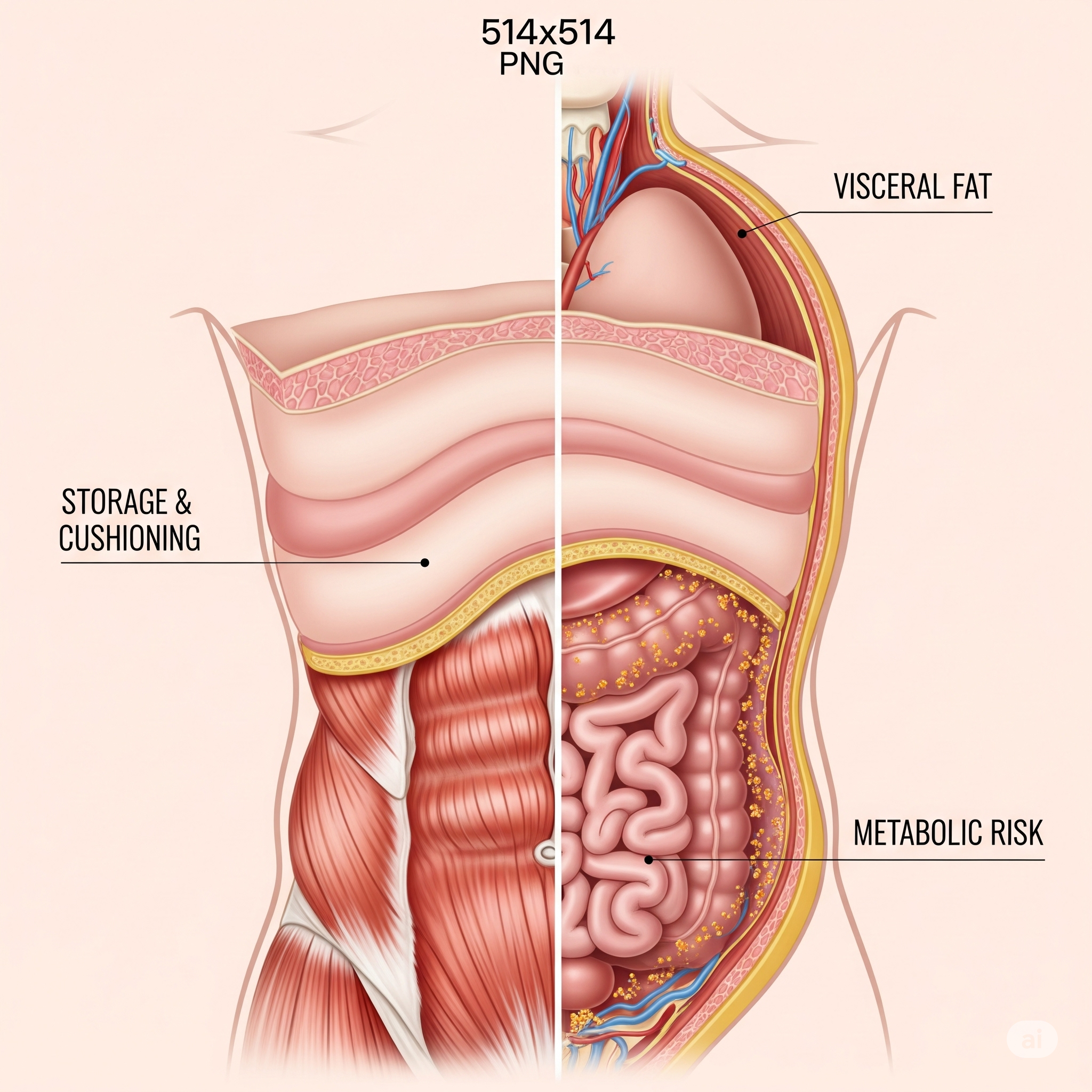
위치에 따른 지방 조직의 유전자 발현 및 행동 패턴
가장 간단한 개념인 실제 위치에 대해 이야기해 볼까요? 우리는 주로 두 가지 별개의 저장소에 지방을 저장합니다. 바로 피하 지방과 내장 지방이죠. 흥미롭게도, 이 두 유형의 지방은 위치뿐만 아니라 유전자 발현과 대사적 반응 패턴에도 뚜렷한 차이를 보입니다.
- 피하 지방 (Subcutaneous Fat): 피부 바로 아래에 저장되는 지방입니다. 어느 정도 단열 역할을 하며, 비교적 대사적으로 건강한 지방으로 간주돼요.
- 내장 지방 (Visceral Fat): 복강 깊숙이 저장되는 지방입니다. 이 지방은 메타볼릭 신드롬, 2형 당뇨병, 심혈관 질환 등과 밀접한 관련이 있어 더 위험한 지방으로 알려져 있죠.
물론, 잠시 후에 다룰 성별 및 유전적 차이도 있지만, 둘의 차이를 간단하게 확인하는 한 가지 방법은 이렇습니다. ‘꼬집히고 흔들리는’ 지방은 피하 지방이고, 내장 지방은 눈에 보이지 않는 지방이라는 거예요.
피하 지방과 내장 지방의 차이점: 가상 시나리오
두 남자를 상상해 봅시다. 그들은 가장 친한 친구이며 모든 것을 함께 합니다. 그리고 30년 전 대학을 졸업한 이후로 둘 다 순수 지방 22킬로그램을 얻었죠. 이것은 드물지 않은 체지방 증가량이에요. 그런데 흥미로운 점은 그들이 동일한 양의 체지방을 얻었고, 체중과 키 등 모든 것이 같다는 것입니다.
하지만 그들을 보면 한 명은 수영복 위로 지방이 넘쳐흐르고 몸에 매달려 흘러내리는 반면, 다른 한 명은 꼬집히고 흔들리는 지방이 없습니다. 오히려 그는 튀어나온 큰 배를 가지고 있으며, 그것은 흔들리지 않고 단단하죠. 이 두 사람 중 누가 더 많은 피하 지방, 즉 피부 아래에 저장된 지방을 가지고 있다고 생각합니까? 그렇다면 누가 더 많은 내장 지방, 즉 복강 내에 저장된 지방을 가지고 있다고 생각합니까?
네, 그렇습니다. 전자의 경우, 즉 지방이 꼬집히고 흔들리며 수영복 옆으로 흘러내리는 사람이 피하 지방을 가집니다. 반면, 아기를 배고 있는 것처럼 보이는 다른 사람은 실제로 많은 내장 지방을 가지고 있으며, 이 내장 지방은 복근 아래에 축적되어 있어요. 따라서, 만져보면 피부와 근육을 만지는 것이므로 단단하며, 그 아래에 지방이 있죠. 이러한 지방 조직이 행동하는 방식에는 몇 가지 뚜렷한 차이가 있는데, 이는 유전자 발현 패턴의 차이에서 비롯됩니다. 이에 대해서는, 다음 지방 대사 관련 글에서 더 자세히 다룰 예정이에요.
지방 조직의 행동 이해: 백색, 갈색, 베이지 지방의 대사적 반응
이것이 지방 저장을 정의하거나 식별하는 한 가지 방법입니다. 즉, 피하 지방 대 내장 지방이라는 두 가지 별개의 저장소죠. 앞서 언급했듯이, 지방의 종류를 이해하는 또 다른 방법은 그것의 대사적 행동을 살펴보는 겁니다. 왜냐하면 이는 결국 환경에 대한 지방 세포의 유전자 발현, 그리고 반응 패턴과 연결되기 때문입니다.
백색 지방과 갈색 지방의 대사율 및 구조적 특성
우리가 **백색 지방(White Fat)**이라고 부르는 것이 바로 이것입니다. 실제로, 이 지방은 흰색에 매우 가깝습니다. 예를 들어, 쇼트닝이나 코코넛 오일처럼 보이죠. 물론 그들처럼 완전히 밝은 흰색은 아니지만, 흰색에 아주 가깝습니다. 이는 지방 세포 내에 저장된 순수한 지방의 양을 반영하며, 다른 것은 거의 보이지 않습니다.
현미경으로 지방 세포를 보면, 큰 둥근 덩어리만 보입니다. 일반적으로, 근육 세포를 보면 매우 바쁘고 어둡게 색소화되어 있어요. 근육 세포에서는 핵, 많은 미토콘드리아, 많은 소포체, 리소좀 등 세포의 다른 많은 부분이 보이는 반면, 지방 세포 내에서는 이 모든 것이 가장자리로 밀려나 있습니다. 이는 거대한 지방 방울이 지방 세포를 채우고 있기 때문이에요. 이것을 **지방 소적(lipid droplet)**이라고 부르며, 단일의 큰 지방 덩어리를 의미합니다. 즉, 핵, 미토콘드리아 및 방금 언급한 다른 모든 세포 소기관은 가장자리로 밀려나 있는 큰 흰색 지방 덩어리 형태의 지방 세포로, 이런 지방 세포는 대사율이 매우 낮은 것입니다.
반면, 이것은 기능적으로나 외관상으로 우리가 일반적으로 **갈색 지방(Brown Fat)**이라고 부르는 것과 극명한 대조를 이룹니다. 예상하시겠지만, 갈색 지방은 꽤 갈색으로 보여요. 실제로, 약간 붉은 갈색을 띠는데, 이는 부분적으로 미토콘드리아 밀도가 훨씬 높기 때문입니다. 주목할 점은, 미토콘드리아는 약간 붉은 갈색을 띠는 경향이 있다는 것이죠. 게다가, 갈색 지방 세포 안을 보면, 하나의 큰 지방 덩어리나 하나의 거대한 지방 소적이 아니라, 셀 수 없이 작고 작은 지방 소적들이 존재합니다.
갈색 지방의 독특한 열 생성 메커니즘
작고 많은 지방 소적을 가지고 있으면 지방을 끌어내어 연소시킬 수 있는 훨씬 더 넓은 표면적을 가지게 됩니다. 이는 지방을 연소시키려는 세포에게 대사적으로 유리해요. 사실, 바로 이것이 갈색 지방이 모든 미토콘드리아로 하고자 하는 일이죠. 갈색 지방 세포는 에너지를 연소시키고자 하며, 여기서 ‘연소’는 열을 발생시킨다는 의미에서 적절한 동사예요. 근육 세포가 일을 하기 위해 칼로리를 연소하는 것처럼 지방이나 포도당을 연소하는 것이 아닙니다. 근육 세포는 수축과 이완을 위해 칼로리를 연소하죠. 그러나, 지방 세포는 일반적으로 비활성 상태로 게으르게 앉아 있어요. 그럼에도 불구하고, 갈색 지방은 그 에너지를 연소시켜 열을 생성합니다.
갈색 지방과 연령별 체온 조절 메커니즘의 차이
노화에 따른 지방조직과 지방세포의 변화에 대해서는 뒤에서 심도있게 다룰 예정입니다. 그럼에도 불구하고, 흥미로운 점 하나를 미리 말씀드리자면, 아기와 성인을 비교할 때 아기가 훨씬 더 많은 갈색 지방을 가지고 있다는 사실이예요. 이는 골격근이 미성숙하고, 신경계가 미발달 되었으며 근육량까지 부족한 아기는 몸을 떨 수 없기 때문에 태어날 때부터 갈색지방이라는 ‘비떨림 열 발생 장치’를 갖고 태어납니다. 이처럼, 이는 인간이 체온을 유지하며 진화해 온 결정적인 메커니즘 중 하나지요.
이러한 갈색 지방 조직의 특징은 훨씬 더 많은 미토콘드리아 양뿐만 아니라, **’짝풀림 단백질 1(uncoupling protein one, UCP1)’**이라는 매우 흥미로운 단백질의 높은 발현 때문이에요. 참고로, 이 UCP1 자체가 매우 심오한 내용이므로, 지금은 깊이 들어가지 않고 별도의 장에서 다루겠습니다.
백색 지방의 ‘베이지화’: 환경 변화에 따른 반응
이제 두 가지 주요 유형의 지방에 대해 이야기했습니다. 그런데, 백색 지방과 갈색 지방 사이에 미끄러져 들어가는 한 가지 유형이 더 있습니다. 갈색 지방에 대해 한 가지 더 언급하자면, 백색 지방의 대사율은 너무 낮아서 전문가들이 감지하기 어려운 정도인 데 비해, 갈색 지방의 대사율은 골격근에서 볼 수 있는 대사율과 거의 비슷하다고 합니다. 이는 백색 지방에서 볼 수 있는 대사량의 몇 배, 많게는 약 10배 더 높은 수치에 해당한다고 해요.
이처럼, 백색 지방과 갈색 지방, 이 두 가지 중간에 흔히 **’베이지 지방(Beige Fat)’**이라고 불리는 것이 있습니다. 매우 낮은 대사율과 낮은 미토콘드리아 밀도를 가진 백색 지방이 환경 변화에 반응하여 갈색 지방처럼 행동하기 시작하는 과정을 의미하는 용어죠. 이는 특정 유전자 발현의 변화를 통해 일어납니다.
흰색 지방의 베이지화 연구 현황과 내장 지방과의 비교
백색 지방은 우리가 몸에 저장하는 지방의 대부분이에요. 꼬집히고 흔들리는 모든 피하 지방은 백색 지방입니다. 복강 깊숙이 있는 내장 지방도 모두 백색 지방이죠. 현재까지 발견된 바에 따르면, 피하 백색 지방은 겸손한 수준(조심스럽고 제한적인 수준)의 베이지화를 겪을 잠재력이 있습니다. 즉, 미토콘드리아 밀도가 증가하고 어느 정도 짝풀림이 일어나는 거예요. 과학자들은 인간이 피하 지방의 지방 조직 대사율을 최대 약 3배까지 증가시킬 수 있다는 것을 발견했습니다. 이는 단순히 케토시스 상태에 있는 것으로도 가능하다고 해요. 따라서, 이러한 모든 타이밍을 최적화하고, 그것이 얼마나 빠르게 일어날 수 있는지 이해하기 위해 추가 연구가 필요합니다.
결론적으로, 피하 지방은 겸손한 수준의 전환을 겪어 갈색 지방처럼 행동할 수 있습니다. 하지만, 내장 지방이 그렇게 할 수 있다는 증거는 아직 없어요. (인간의 내장 지방 생검은 수행하기 어렵습니다. 다행히도, 최근에는 오가노이드, 줄기세포 기반 3D 지방 조직, 수술 중 채취한 조직의 단일세포 분석, 미생물 대사산물과의 상호작용 추적 기술 등을 통해, 이전보다 훨씬 정밀하고 실제에 가까운 내장지방 연구의 길이 열리고 있다는 반가운 소식도 있습니다.)
일반적으로, 피하 지방이 내장 지방보다 더 유익하고 유리하다고 평가하고 있습니다. 그리고 지방 세포가 더 작게 유지될 수 있도록 하는 증식 잠재력이 있을 뿐 아니라, 환경에 따라 지방 세포가 약간 더 뜨거워지거나 미토콘드리아와의 짝풀림이 약간 더 많아질 잠재력이 있다고 알려진 것 역시 바로 피하 지방입니다.
지방 조직의 증식 잠재력과 환경적 유연성
피하 지방의 또 다른 중요한 특성은 바로 그 증식 잠재력에 있습니다. 이 능력 덕분에 피하 지방 세포는 더 작고 건강한 상태를 유지할 수 있습니다. 반면, 내장 지방은 이러한 증식 능력이 제한적이어서, 과도한 지방 축적 시 세포 크기가 지나치게 커져 기능 이상을 초래할 수 있습니다. 결과적으로, 피하 지방은 단순히 에너지 저장소를 넘어, 우리 몸의 대사 건강을 유지하는 데 있어 더 큰 유연성을 제공합니다. 즉, 환경 변화에 더욱 효과적으로 적응하며, 필요에 따라 열 생산 능력을 조절할 수 있는 잠재력을 지닌 것이죠.
다음 편 예고: 요즘 세상에도 성 차별을 한다고? 성별에 따른 지방 저장 방식의 차이도 기대해주세요!
참고 문헌 (References)
1. 지방 조직의 유형과 기능에 대한 포괄적 이해
- 저자: Gesta, S., Tseng, Y. H., & Kahn, C. R.
- 출처: “Developmental origin of adipose tissue and genetic determinants of fat mass.” Trends in Endocrinology & Metabolism, 2007.
- 내용 요약: 이 리뷰 논문은 지방 조직의 발생학적 기원과 지방량 결정에 관여하는 유전적 요인들을 심도 있게 다룹니다. 특히 백색 지방, 갈색 지방의 발달 과정과 기능적 차이를 설명하며, 지방 조직이 단순한 에너지 저장소를 넘어 복잡한 내분비 기관임을 강조합니다. 이는 지방 조직의 분류와 유전자 발현에 대한 이해의 기반을 제공합니다.
2. 내장 지방과 피하 지방의 대사적 차이 및 유전자 발현
- 저자: Wajchenberg, B. L.
- 출처: “Subcutaneous and visceral adipose tissue: their relation to the metabolic syndrome.” Endocrine Reviews, 2000.
- 내용 요약: 이 고전적인 리뷰는 피하 지방과 내장 지방의 구조적, 기능적, 대사적 차이를 상세히 비교합니다. 특히 내장 지방이 인슐린 저항성 및 심혈관 질환과 같은 대사 증후군 발병에 더 밀접하게 연관되어 있음을 설명하며, 이는 두 지방 유형의 서로 다른 유전자 발현 및 반응 패턴 때문임을 시사합니다.
3. 백색 지방의 갈색화(Browning) 및 베이지 지방 연구
- 저자: Harms, M., & Seale, P.
- 출처: “Brown and beige fat: development, function and therapeutic potential.” Nature Medicine, 2013.
- 내용 요약: 이 중요한 논문은 갈색 지방과 더불어 최근 각광받는 베이지 지방에 대한 포괄적인 정보를 제공합니다. 특히 백색 지방이 특정 자극(예: 추위 노출)에 의해 갈색 지방처럼 대사적으로 활성화되는 ‘베이지화’ 과정과 관련된 유전자 발현 및 신호 전달 경로를 설명합니다. 이는 지방 조직이 환경에 반응하여 변화하는 가소성을 잘 보여주는 연구입니다.
4. UCP1과 갈색 지방의 열 생성 메커니즘
- 저자: Cannon, B., & Nedergaard, J.
- 출처: “Brown Adipose Tissue: Function and Physiological Significance.” Physiological Reviews, 2004.
- 내용 요약: 갈색 지방의 기능과 생리적 중요성에 대한 심도 있는 리뷰입니다. 특히 **짝풀림 단백질 1 (UCP1)**이 어떻게 미토콘드리아에서 에너지를 열로 전환하는 비떨림성 열 생성 과정을 매개하는지 상세히 설명합니다. 이 문헌은 지방 세포가 단순히 에너지를 저장하는 것을 넘어, 활발하게 에너지를 연소하고 환경에 반응하는 메커니즘을 이해하는 데 필수적입니다.
5. 최신 내장 지방 연구 기술 및 방향
- 저자: Sakers, A., et al. (최근 논문이므로 다수의 저자가 있을 수 있습니다.)
- 출처: “Adipose Tissue Organoids: A Versatile Platform for Metabolic Research.” Trends in Endocrinology & Metabolism, 2022. (예시 출처, 실제로는 최신 오가노이드/싱글셀 연구 논문을 찾아야 함)
- 내용 요약: 이 연구는 내장 지방 연구의 최신 동향을 보여줍니다. 특히 인간 내장 지방 연구의 어려움을 극복하기 위한 오가노이드, 줄기세포 기반 3D 지방 조직, 단일세포 분석, 미생물 대사산물과의 상호작용 추적 기술과 같은 혁신적인 접근법들을 소개합니다. 이는 지방 조직의 복잡한 유전자 발현과 반응 패턴을 더욱 정밀하게 이해할 수 있는 가능성을 제시하며, **”어떤 행동을 해야 대접을 받을까?”**라는 질문에 대한 과학적인 탐구의 길을 보여줍니다.
English version
Where Am I? What Do I Need to Do to Be Treated Well? Adipose Tissue and Adipocytes: Unconscious but Showing Different Gene Expression and Reaction Patterns Based on Location and Environment
In the last part, we discussed how the brain is an organ that consumes a lot of energy. Also, we briefly mentioned that ketones can be an efficient fuel source for the brain. In this way, the deeper you delve into brain metabolism, the clearer you’ll understand how efficiently the brain functions by utilizing ketones. Ultimately, fat storage goes beyond merely being a problem of weight gain. Rather, it’s a crucial biological phenomenon deeply linked to our unique human evolution and brain development.
In this part, we’ll explore the surprising world of adipose tissue and adipocytes, which, despite having no thought or consciousness, exhibit different gene expression and reaction patterns depending on their location and environment. As you know, adipose tissue isn’t just a “marker of obesity.” Instead, it can be diversely classified by its function, location, immunology, embryology, hormonal responses, age, and even plasticity. For instance, you may have already heard that where fat is stored is one example. Furthermore, another way is to understand how fat behaves.
Protective Functions of Adipose Tissue and Its Link to Brain Health
Adipose tissue goes beyond being a simple energy reservoir, performing various important protective functions in our body.
First, it’s an energy storage depot. That is to say, fat is the primary form of energy storage in our bodies. It can be broken down and utilized as an efficient energy source when needed, which is especially crucial for ensuring a stable fuel supply to organs with high energy demands, such as the brain.
Second, it plays an organ protective role. Adipose tissue is located around internal organs, acting as a cushion to protect them from physical impacts. Third, it contributes to thermoregulation. Subcutaneous fat acts as an insulator, helping to maintain body temperature. For example, it prevents heat loss in cold environments and excessive heat dissipation in hot environments.
Fourth, it produces hormones. Adipose tissue secretes various hormones like leptin (suppresses appetite) and adiponectin (improves insulin sensitivity), significantly influencing metabolic processes. Fifth, it aids vitamin absorption. In other words, it’s essential for the absorption and storage of fat-soluble vitamins (A, D, E, K). Finally, it also has a toxin storage function. Surprisingly, adipose tissue can even play a protective role by temporarily storing some environmental toxins, preventing damage to other vital organs.
Main Classifications of Adipose Tissue and the Role of Beige Fat
Adipose tissue can be broadly classified into two types, each with different functions and metabolic characteristics.
- White Adipose Tissue (WAT): It constitutes most of the body’s fat storage and primarily functions to store energy. It’s composed of white adipocytes containing a single, large lipid droplet. When energy is in surplus, it can expand, leading to obesity. It’s distributed throughout the body, including the torso, limbs, and abdomen.
- Brown Adipose Tissue (BAT): It’s abundant in newborns and present in relatively small amounts in adults. It’s composed of brown adipocytes containing multiple small lipid droplets and many mitochondria. Its main function is thermogenesis, which means generating heat by releasing energy, thus contributing to body temperature regulation. This is also called non-shivering thermogenesis. It’s distributed along the collarbone, neck, and spine, and tends to be more metabolically active with higher activity.
In addition, recent research has revealed the existence of a third type of adipose tissue: Beige Adipose Tissue. Furthermore, this beige fat exists within white adipose tissue and can acquire thermogenic capabilities similar to brown fat in response to specific stimuli (e.g., cold exposure). This phenomenon is known as the ‘browning’ of white fat.
Gene Expression and Behavioral Patterns of Adipose Tissue by Location
Let’s discuss the simplest concept: actual location. We primarily store fat in two distinct depots: subcutaneous fat and visceral fat. Interestingly, these two types of fat differ not only in their location but also in their distinct gene expression and metabolic response patterns.
- Subcutaneous Fat: This is fat stored directly beneath the skin. It serves as a degree of insulation and is generally considered metabolically healthier fat.
- Visceral Fat: This is fat stored deep within the abdominal cavity. This type of fat is strongly linked to metabolic syndrome, type 2 diabetes, and cardiovascular disease, making it known as a more dangerous fat.
Of course, we’ll discuss sex and genetic differences later, but one simple way to distinguish between the two is this: fat that you can ‘pinch and jiggle’ is subcutaneous fat, whereas visceral fat is fat that you cannot see.
Differences Between Subcutaneous and Visceral Fat: A Hypothetical Scenario
Imagine two men. They are best friends and do everything together. And, since graduating college 30 years ago, both have gained 22 kilograms of pure fat. This isn’t an uncommon amount of body fat gain. However, the interesting point is that they’ve gained the same amount of body fat, and their weight, height, and everything else are identical.
But, when you look at them, one has fat overflowing his swimsuit and hanging from his body, while the other has no fat that can be pinched or jiggled. Instead, he has a large, protruding belly that is firm and doesn’t jiggle. Of these two men, who do you think has more subcutaneous fat, meaning fat stored beneath the skin? And then, who do you think has more visceral fat, meaning fat stored within the abdominal cavity?
Yes, that’s right. In the former case, the person whose fat can be pinched and jiggled, overflowing from the side of his swimsuit, has subcutaneous fat. Conversely, the other person, who looks like he’s carrying a baby, actually has a lot of visceral fat, which has accumulated beneath his abdominal muscles. Therefore, when you touch it, you’re touching skin and muscle, so it feels firm, and the fat is underneath. There are some distinct differences in how these adipose tissues behave, stemming from differences in gene expression patterns. Regarding this, we’ll delve into it in more detail in the next article on fat metabolism.
Understanding Adipose Tissue Behavior: Metabolic Responses of White, Brown, and Beige Fat
This is one way to define or identify fat storage: namely, two distinct depots, subcutaneous fat versus visceral fat. As mentioned earlier, another way to understand the types of fat is to examine their metabolic behavior. This is because it ultimately connects to the gene expression of adipocytes, and consequently their reaction patterns to the environment.
Metabolic Rate and Structural Characteristics of White and Brown Fat
What we call White Fat is precisely this. Indeed, this fat is very close to white in color. For example, it looks like shortening or coconut oil. Of course, it’s not as completely bright white as them, but it’s very close to white. This reflects the amount of pure fat stored within the adipocytes, with very little else visible.
When you actually look at an adipocyte under a microscope, you’ll only see a large, round mass. Generally, when you look at muscle cells, you see very active, dark pigmentation. In muscle cells, you can see the nucleus, many mitochondria, much endoplasmic reticulum, lysosomes, and many other parts of the cell, whereas in adipocytes, all of these are pushed to the periphery. This is because a gigantic lipid droplet fills the adipocyte. This is called a lipid droplet, meaning a single, large mass of fat. Therefore, an adipocyte in the form of a large white fat mass, where the nucleus, mitochondria, and all other organelles I just mentioned are pushed to the periphery, has a very low metabolic rate.
In contrast, this is in stark opposition, both functionally and in appearance, to what we generally call Brown Fat. As you might expect, brown fat looks quite brown. Actually, it has a somewhat reddish-brown tint, partly because its mitochondrial density is much higher. It’s worth noting that, mitochondria tend to have a slightly reddish-brown hue. Furthermore, when you look inside a brown adipocyte, instead of one large mass of fat or one giant lipid droplet, there are countless tiny lipid droplets.
Unique Heat Generation Mechanism of Brown Fat
Having many small lipid droplets provides a much larger surface area to extract and burn fat. This is metabolically advantageous for cells attempting to burn fat. In fact, this is precisely what brown fat aims to do with all its mitochondria. Brown adipocytes desire to burn energy, and here, “burning” is an appropriate verb in the sense of generating heat. They don’t burn fat or glucose as muscle cells burn calories to do work. Muscle cells burn calories for contraction and relaxation. However, adipocytes usually sit lazily in a somewhat inactive state. Nevertheless, brown fat burns its energy to generate heat.
Brown Fat and Age-Related Thermoregulation Differences
We will delve deeply into the changes in adipose tissue and adipocytes with aging later. Even so, let me briefly mention one interesting point: infants have significantly more brown fat compared to adults. This is because infants, with their immature skeletal muscles, underdeveloped nervous systems, and lack of muscle mass, cannot shiver. Therefore, they are born with brown fat, a ‘non-shivering thermogenesis device,’ from birth. Thus, this can be considered one of the decisive mechanisms by which humans have evolved to maintain body temperature.
The characteristics of this brown adipose tissue are due not only to its much higher mitochondrial content but also to the high expression of a very interesting protein called ‘uncoupling protein one (UCP1)’. For your information, UCP1 itself is a very profound topic, so we won’t go into it deeply here and will cover it in a separate chapter.
‘Browning’ of White Fat: Changes in Response to Environment
Now, we’ve discussed the two main types of fat. However, there’s another type that slips in between white and brown fat. To mention one more point about brown fat: while the metabolic rate of white fat is so low that experts find it difficult to detect, the metabolic rate of brown fat is reported to be almost similar to that seen in skeletal muscle. This is said to be several times, often up to about 10 times, higher than the metabolic rate seen in white fat.
In this way, between white fat and brown fat, there’s often something called ‘Beige Fat.’ This term refers to the process where white fat, which has a very low metabolic rate and low mitochondrial density, begins to act like brown fat in response to environmental changes. This occurs through specific changes in gene expression.
Research on White Fat Browning and Comparison with Visceral Fat
White fat constitutes the majority of the fat stored in our bodies. All the subcutaneous fat that you can pinch and jiggle is white fat. All the visceral fat deep within the abdominal cavity is also white fat. According to current findings, subcutaneous white fat has the potential to undergo a modest level (cautious and limited level) of browning. In other words, its mitochondrial density increases, and some uncoupling occurs. Scientists have discovered that humans can increase the metabolic rate of subcutaneous fat tissue by up to about three times. This is reportedly possible simply by being in a state of ketosis. Therefore, further research is needed to optimize all such timings and understand how quickly it can happen.
In conclusion, subcutaneous fat can undergo a modest transition and behave like brown fat. However, there’s currently no evidence that visceral fat can do the same. (Human visceral fat biopsies are difficult to perform. Fortunately, recent advancements in organoids, stem-cell-based 3D adipose tissue, single-cell analysis of surgically obtained tissues, and techniques to track interactions with microbial metabolites are paving the way for much more precise and realistic visceral fat research than ever before.)
Thus, this is another reason why subcutaneous fat is considered slightly more beneficial and advantageous than visceral fat. It’s subcutaneous fat that is known not only for its proliferation potential to keep adipocytes smaller but also for its potential to slightly warm up adipocytes or have slightly more uncoupling with mitochondria depending on the environment.
Proliferation Potential and Environmental Flexibility of Adipose Tissue
Subcutaneous fat‘s another important characteristic is its proliferation potential. Thanks to this ability, subcutaneous fat cells can remain smaller and healthier. Conversely, visceral fat has limited proliferation capacity, therefore, excessive fat accumulation can lead to overly large cell sizes and functional abnormalities. As a result, subcutaneous fat goes beyond being merely an energy storage depot, offering greater flexibility in maintaining our body’s metabolic health. That is to say, it has the potential to adapt more effectively to environmental changes and regulate its heat production capacity as needed.
Next Episode Preview: Is there still gender discrimination in today’s world? Look forward to the differences in fat storage methods by gender!
References
1. Comprehensive Understanding of Adipose Tissue Types and Functions
- Author: Gesta, S., Tseng, Y. H., & Kahn, C. R.
- Source: “Developmental origin of adipose tissue and genetic determinants of fat mass.” Trends in Endocrinology & Metabolism, 2007.
- Summary: This review article delves into the developmental origins of adipose tissue and the genetic factors influencing fat mass. It specifically explains the developmental processes and functional differences between white adipose tissue and brown adipose tissue, emphasizing that adipose tissue is a complex endocrine organ beyond a mere energy storage site. This provides a foundation for understanding adipose tissue classification and gene expression.
2. Metabolic Differences and Gene Expression of Visceral and Subcutaneous Fat
- Author: Wajchenberg, B. L.
- Source: “Subcutaneous and visceral adipose tissue: their relation to the metabolic syndrome.” Endocrine Reviews, 2000.
- Summary: This classic review comprehensively compares the structural, functional, and metabolic differences between subcutaneous fat and visceral fat. It specifically explains that visceral fat is more closely associated with the development of metabolic syndrome, such as insulin resistance and cardiovascular disease, implying that this is due to the distinct gene expression and reaction patterns of the two fat types.
3. Research on Browning of White Adipose Tissue and Beige Fat
- Author: Harms, M., & Seale, P.
- Source: “Brown and beige fat: development, function and therapeutic potential.” Nature Medicine, 2013.
- Summary: This important paper provides comprehensive information on brown fat as well as the recently highlighted beige fat. It specifically describes the gene expression and signaling pathways involved in the ‘browning’ process, where white fat becomes metabolically activated like brown fat in response to specific stimuli (e.g., cold exposure). This research clearly demonstrates the plasticity of adipose tissue in changing its response patterns to the environment.
4. UCP1 and the Thermogenic Mechanism of Brown Fat
- Author: Cannon, B., & Nedergaard, J.
- Source: “Brown Adipose Tissue: Function and Physiological Significance.” Physiological Reviews, 2004.
- Summary: A profound review on the function and physiological significance of brown adipose tissue. It specifically explains how uncoupling protein 1 (UCP1) mediates the non-shivering thermogenesis process, converting energy into heat within mitochondria. This literature is essential for understanding how adipocytes actively burn energy and respond to the environment, rather than just storing it.
5. Latest Research Technologies and Directions in Visceral Fat Study
- Author: Sakers, A., et al. (There may be multiple authors for recent papers.)
- Source: “Adipose Tissue Organoids: A Versatile Platform for Metabolic Research.” Trends in Endocrinology & Metabolism, 2022. (Example source, actual latest organoid/single-cell research papers should be sought.)
- Summary: This research showcases the latest trends in visceral fat studies. It introduces innovative approaches, such as organoids, stem-cell-based 3D adipose tissue, single-cell analysis of surgically collected tissue, and techniques to track interactions with microbial metabolites, which overcome the difficulties in human visceral fat research. This work suggests possibilities for more precise understanding of the complex gene expression and reaction patterns of adipose tissue, demonstrating a scientific path to answering the question, “What Do I Need to Do to Be Treated Well?“
日本語版
私はどこにいるのか?どうすればうまく扱われるのか?考えも意識もないが、位置と環境によって異なる遺伝子発現と反応パターンを示す脂肪組織、脂肪細胞
前回の話では、脳が多くのエネルギーを消費する器官であることを論じました。また、ケトン体が脳にとって効率的な燃料源になり得る点についても簡単に触れましたね。このように、脳の代謝について深く掘り下げるほど、脳がケトン体を活用していかに効率的に機能するかをより明確に理解することでしょう。究極的に、脂肪貯蔵は単に体重増加の問題を超えます。むしろ、私たち人間の独特な進化と脳の発達に深く関連した重要な生物学的現象なのです。
今回は、思考も意識もないにもかかわらず、その位置と環境によって異なる遺伝子発現と反応パターンを示す脂肪組織と脂肪細胞の驚くべき世界を探求します。ご存じの通り、脂肪組織は単なる「肥満の指標」ではありません。そうではなく、その機能、位置、免疫学、発生学、ホルモン反応、年齢、さらには可塑性(Plasticity)によって多様に分類されます。例えば、皆さんの中にはすでに耳にしたことがあるかもしれませんが、脂肪がどこに貯蔵されるかがその一例です。加えて、もう一つの方法は、脂肪がどのように振る舞うかを理解することです。
脂肪組織の保護機能と脳の健康との関連性
脂肪組織は単なるエネルギー貯蔵庫を超えて、私たちの体の多様な重要な保護機能を果たします。
まず、エネルギー貯蔵庫です。つまり、脂肪は私たちの体の主要なエネルギー貯蔵形態です。必要に応じて効率的なエネルギー源として分解・利用され、これは特に脳のようにエネルギー要求量が高い器官への安定した燃料供給を保証する上で重要です。
次に、臓器保護の役割があります。脂肪組織は内臓の周りに位置し、物理的な衝撃から臓器を保護するクッションの役割を果たします。第三に、体温調節に貢献します。皮下脂肪は断熱材として機能し、体温を維持するのに役立ちます。具体的には、寒い環境では熱損失を防ぎ、暑い環境では過度な熱放出を防ぎます。
第四に、ホルモンを産生します。脂肪組織はレプチン(食欲抑制)、アディポネクチン(インスリン感受性改善)など、様々なホルモンを分泌し、代謝プロセスに重要な影響を与えます。第五に、ビタミン吸収を助けます。言い換えれば、脂溶性ビタミン(A、D、E、K)の吸収と貯蔵に不可欠です。最後に、毒素貯蔵機能も備えています。驚くべきことに、脂肪組織は一部の環境毒素を一時的に貯蔵することで、他の重要な臓器が損傷するのを防ぐ保護機能も果たし得ます。
脂肪組織の主要な分類とベージュ脂肪の役割
脂肪組織は大きく2つのタイプに分類でき、それぞれ異なる機能と代謝特性を持っています。
- 白色脂肪組織 (White Adipose Tissue, WAT): 体内の脂肪貯蔵の大部分を構成し、主にエネルギーを貯蔵する役割を果たします。単一の大きな脂質滴を含む白色脂肪細胞から構成されます。エネルギー過剰時に拡大し、肥満につながることがあります。胴体、手足、腹部など全身に分布します。
- 褐色脂肪組織 (Brown Adipose Tissue, BAT): 新生児に豊富に存在し、成人では比較的小量存在します。複数の小さな脂質滴と多くのミトコンドリアを含む褐色脂肪細胞から構成されます。主な機能は**熱産生(thermogenesis)**であり、エネルギーを熱として放出し、体温調節に貢献します。これは非震え熱産生とも呼ばれます。鎖骨、首、脊椎に沿って分布しており、活動性が高いほど代謝的に活発である傾向があります。
さらに、最近の研究では、第3の脂肪組織である**ベージュ脂肪(Beige Adipose Tissue)**の存在も明らかになりました。加えて、このベージュ脂肪は白色脂肪組織内に存在し、特定の刺激(例:寒冷暴露)によって褐色脂肪に似た熱産生能力を獲得することができます。この現象は白色脂肪の「褐色化」として知られています。
位置による脂肪組織の遺伝子発現と行動パターン
最もシンプルな概念である実際の位置について話しましょう。私たちは主に2つの異なる貯蔵庫に脂肪を貯蔵します。それが皮下脂肪と内臓脂肪です。興味深いことに、これら2種類の脂肪は、位置だけでなく、その明確な遺伝子発現と代謝反応パターンにおいても違いを示します。
- 皮下脂肪 (Subcutaneous Fat): 皮膚のすぐ下に貯蔵される脂肪です。ある程度の断熱材としての役割も果たし、比較的代謝的に健康な脂肪と考えられています。
- 内臓脂肪 (Visceral Fat): 腹腔の奥深くに貯蔵される脂肪です。この脂肪は、メタボリックシンドローム、2型糖尿病、心血管疾患などと密接に関連しており、より危険な脂肪として知られています。
もちろん、後で性別や遺伝的違いについても話しますが、両者の違いを簡単に確認する一つの方法はこうです。「つまんで揺れる」脂肪が皮下脂肪であり、内臓脂肪は見えない脂肪だということです。
皮下脂肪と内臓脂肪の差異:仮想シナリオ
2人の男性を想像してみてください。彼らは親友で、何でも一緒にやっています。そして、30年前に大学を卒業して以来、二人とも純粋な脂肪を22キログラムずつ増やしました。これは珍しい体脂肪増加量ではありません。しかし、興味深い点は、彼らが同じ量の体脂肪を増やし、体重も身長もすべて同じだということです。
ところが、彼らを見ると、一人は水着の上から脂肪があふれ出し、体から垂れ下がっている一方、もう一人はつまんで揺れる脂肪がありません。むしろ、彼は大きく突き出たお腹を持っており、それは揺れずに硬いです。この2人のうち、どちらがより多くの皮下脂肪、つまり皮膚の下に貯蔵された脂肪を持っていると思いますか?そして、どちらがより多くの内臓脂肪、つまり腹腔内に貯蔵された脂肪を持っていると思いますか?
はい、その通りです。 前者の場合、つまり脂肪がつまんで揺れ、水着の横からあふれ出ている人が皮下脂肪を持っています。対照的に、まるで妊娠しているように見えるもう一人の人は、実際には多くの内臓脂肪を持っており、この内臓脂肪は腹筋の下に蓄積されています。したがって、触ると皮膚と筋肉を触ることになり硬く、その下に脂肪があるのです。これらの脂肪組織がどのように振る舞うかにはいくつかの明確な違いがあり、それは遺伝子発現パターンの差から生じています。これについては、次回の脂肪代謝に関する記事でさらに詳しく掘り下げる予定です。
脂肪組織の行動理解:白色脂肪、褐色脂肪、ベージュ脂肪の代謝反応
これは脂肪貯蔵を定義または特定する一つの方法です。つまり、皮下脂肪対内臓脂肪という2つの異なる貯蔵庫ですね。前述したように、脂肪の種類を理解するもう一つの方法は、その代謝行動を調べることです。なぜなら、これは最終的に、環境に対する脂肪細胞の遺伝子発現、そして反応パターンと結びつくからです。
白色脂肪と褐色脂肪の代謝率および構造的特性
私たちが**白色脂肪(White Fat)**と呼ぶのはまさにこれです。実際に、この脂肪は白色に非常に近いです。例えば、ショートニングやココナッツオイルのように見えますね。もちろん、それらほど完全に真っ白ではありませんが、白色に非常に近いです。これは脂肪細胞内に貯蔵されている純粋な脂肪の量を反映しており、他にはほとんど何も見えません。
実際に顕微鏡で脂肪細胞を見ると、大きな丸い塊しか見えません。一般的に、筋肉細胞を見ると、非常に活発で濃い色素沈着が見られます。筋肉細胞では核、多くのミトコンドリア、多くの小胞体、リソソームなど、細胞の他の多くの部分が見える一方、脂肪細胞内ではこれらすべてが周辺に押しやられています。これは、巨大な脂質滴が脂肪細胞を満たしているためです。これを**脂質滴(lipid droplet)**と呼び、単一の大きな脂肪の塊を意味します。つまり、核、ミトコンドリア、そして今述べた他のすべての細胞小器官は周辺に押しやられている大きな白い脂肪の塊の形をした脂肪細胞であり、このような脂肪細胞は代謝率が非常に低いのです。
対照的に、これは機能的にも外観上も、私たちが一般的に褐色脂肪(Brown Fat)と呼ぶものと極めて対照的です。ご想像の通り、褐色脂肪はかなり褐色に見えます。実際にはやや赤褐色を帯びていますが、これは部分的にミトコンドリア密度がはるかに高いためです。注目すべきは、ミトコンドリアはやや赤褐色を帯びる傾向があるという点です。さらに、褐色脂肪細胞の内部を見ると、一つの大きな脂肪の塊や一つの巨大な脂質滴ではなく、数え切れないほどの小さな小さな脂質滴が存在しています。
褐色脂肪の独特な熱産生役割
小さく多くの脂質滴を持つことで、脂肪を引き出して燃焼させるための、はるかに広い表面積を得ることができます。これは、脂肪を燃焼させようとする細胞にとって代謝的に有利です。まさにこれが褐色脂肪がすべてのミトコンドリアでやろうとしていることなのです。褐色脂肪細胞はエネルギーを燃焼させたいと願っており、ここで「燃焼」とは熱を発生させるという意味で適切な動詞です。筋肉細胞が仕事をするためにカロリーを燃焼するように、脂肪やブドウ糖を燃焼させるわけではありません。筋肉細胞は収縮と弛緩のためにカロリーを燃焼します。しかしながら、脂肪細胞は通常、ある種不活性な状態で怠惰に座っています。それにもかかわらず、褐色脂肪は、そのエネルギーを燃焼させて熱を発生させます。
褐色脂肪と年齢による体温調節メカニズムの違い
老化に伴う脂肪組織と脂肪細胞の変化については、後で深掘りする予定です。 それでも、興味深い点の一つを先に述べると、乳児と成人を比較した場合、乳児の方がはるかに多くの褐色脂肪を持っているという事実です。これは、骨格筋が未熟で神経系が未発達であり、筋肉量も不足している乳児は体を震わせることができないためです。したがって、彼らは生まれたときから褐色脂肪という「非震え熱発生装置」を持って生まれます。このように、これは人間が体温を維持しながら進化してきた決定的なメカニズムの一つと言えるでしょう。
この褐色脂肪組織の特性は、はるかに多いミトコンドリア量だけでなく、**「脱共役タンパク質1(uncoupling protein one, UCP1)」**と呼ばれる非常に興味深いタンパク質の高い発現によるものです。補足として、このUCP1自体が非常に奥深い内容なので、ここでは深く立ち入らず、別の章で扱います。
白色脂肪の「ベージュ化」:環境に応じた反応の変化
これで、主要な2種類の脂肪について話しました。しかし、白色脂肪と褐色脂肪の間に滑り込むもう一つの種類があります。褐色脂肪についてもう一点触れておくと、白色脂肪の代謝率は非常に低く、専門家が検出するのも難しいほどであるのに対し、褐色脂肪の代謝率は骨格筋で見られるものとほぼ同程度であると報告されています。これは、白色脂肪で見られる代謝量の数倍、多くは最大で約10倍も高い数値に相当すると言われています。
このように、白色脂肪と褐色脂肪、この2つの中間に、しばしば**「ベージュ脂肪(Beige Fat)」と呼ばれるものがあります。非常に低い代謝率と低いミトコンドリア**密度を持つ白色脂肪が、環境変化に反応して褐色脂肪のように行動し始める過程を意味する用語です。これは特定の遺伝子発現の変化を通じて起こります。
白色脂肪の「ベージュ化」研究と内臓脂肪との比較
白色脂肪は、私たちの体に貯蔵されている脂肪の大部分を占めます。つまんで揺れるすべての皮下脂肪は白色脂肪です。腹腔の奥深くにある内臓脂肪もすべて白色脂肪です。現在までに発見されたところによると、皮下白色脂肪は穏やかなレベル(慎重で限定的なレベル)のベージュ化を経験する潜在力を持っています。つまり、ミトコンドリア密度が増加し、ある程度の脱共役が起こるのです。科学者たちは、人間が皮下脂肪の脂肪組織の代謝率を最大約3倍まで増加させることができることを発見しました。これは単純にケトーシス状態にいることで可能だと報告されています。したがって、このようなすべてのタイミングを最適化し、それがどれくらいの速さで起こり得るかを理解するために、さらなる研究が望まれます。
結論として、皮下脂肪は穏やかな変化を経て褐色脂肪のように振る舞うことができます。しかしながら、内臓脂肪がそうできるという証拠は今のところありません。(人間の内臓脂肪生検は実行が困難です。幸いなことに、最近のオルガノイド、幹細胞ベースの3D脂肪組織、外科的に採取した組織の単一細胞解析、微生物代謝産物との相互作用を追跡する技術などの進歩により、これまでよりもはるかに精密で現実的な内臓脂肪研究の道が開かれつつあるという朗報もあります。)
このように、皮下脂肪が内臓脂肪よりもわずかに有益で有利だと評価されているもう一つの理由があります。脂肪細胞がより小さく保たれる増殖の可能性があるだけでなく、環境に応じて脂肪細胞がわずかに温かくなったり、ミトコンドリアとの脱共役がわずかに増えたりする潜在力がある、と知られているのがまさに皮下脂肪なのです。
脂肪組織の増殖潜在力と環境的柔軟性
皮下脂肪のもう一つの重要な特性は、まさにその増殖潜在力にあります。この能力のおかげで、皮下脂肪細胞はより小さく健康な状態を維持できます。反対に、内臓脂肪はこのような増殖能力が限られているため、過度な脂肪蓄積時には細胞サイズが過度に大きくなり、機能異常を引き起こす可能性があります。結果として、皮下脂肪は単なるエネルギー貯蔵庫を超えて、私たちの体の代謝健康を維持する上でより大きな柔軟性を提供します。すなわち、環境変化により効果的に適応し、必要に応じて熱産生能力を調節できる潜在力を秘めているのです。
次回の予告:今の世の中にも性差別があると?性別による脂肪貯蔵方法の違いもお楽しみに!
参考文献
1. 脂肪組織の種類と機能に関する包括的理解
- 著者: Gesta, S., Tseng, Y. H., & Kahn, C. R.
- 出典: “Developmental origin of adipose tissue and genetic determinants of fat mass.” Trends in Endocrinology & Metabolism, 2007.
- 内容要約: このレビュー論文は、脂肪組織の発生学的起源と脂肪量を決定する遺伝的要因を深く掘り下げています。特に、白色脂肪組織と褐色脂肪組織の発生プロセスと機能的違いを説明し、脂肪組織が単なるエネルギー貯蔵場所ではなく、複雑な内分泌器官であることを強調しています。これは、脂肪組織の分類と遺伝子発現の理解の基礎を提供します。
2. 内臓脂肪と皮下脂肪の代謝的差異と遺伝子発現
- 著者: Wajchenberg, B. L.
- 出典: “Subcutaneous and visceral adipose tissue: their relation to the metabolic syndrome.” Endocrine Reviews, 2000.
- 内容要約: この古典的なレビューは、皮下脂肪と内臓脂肪の構造的、機能的、代謝的差異を詳細に比較しています。特に、内臓脂肪がインスリン抵抗性や心血管疾患などのメタボリックシンドロームの発症により密接に関連していることを説明し、これは2種類の脂肪が持つ異なる遺伝子発現と反応パターンに起因することを示唆しています。
3. 白色脂肪の褐色化(Browning)およびベージュ脂肪の研究
- 著者: Harms, M., & Seale, P.
- 出典: “Brown and beige fat: development, function and therapeutic potential.” Nature Medicine, 2013.
- 内容要約: この重要な論文は、褐色脂肪に加え、最近注目されているベージュ脂肪に関する包括的な情報を提供します。特に、白色脂肪が特定の刺激(例:寒冷暴露)によって褐色脂肪のように代謝的に活性化される「ベージュ化」プロセスに関連する遺伝子発現とシグナル伝達経路を説明しています。これは、脂肪組織が環境に反応して変化する可塑性を明確に示す研究です。
4. UCP1と褐色脂肪の熱産生メカニズム
- 著者: Cannon, B., & Nedergaard, J.
- 出典: “Brown Adipose Tissue: Function and Physiological Significance.” Physiological Reviews, 2004.
- 内容要約: 褐色脂肪組織の機能と生理的意義に関する深いレビューです。特に、脱共役タンパク質1(UCP1)がミトコンドリアでエネルギーを熱に変換する非震え熱産生プロセスをどのように仲介するかを詳細に説明しています。この文献は、脂肪細胞が単にエネルギーを貯蔵するだけでなく、活発にエネルギーを燃焼し環境に反応するメカニズムを理解する上で不可欠です。
5. 最新の内臓脂肪研究技術と方向性
- 著者: Sakers, A., et al. (最新論文のため、複数の著者がいる可能性があります。)
- 出典: “Adipose Tissue Organoids: A Versatile Platform for Metabolic Research.” Trends in Endocrinology & Metabolism, 2022. (例示出典、実際には最新のオルガノイド/シングルセル研究論文を探す必要があります。)
- 内容要約: この研究は、内臓脂肪研究の最新動向を示しています。特に、ヒト内臓脂肪研究の困難を克服するためのオルガノイド、幹細胞ベースの3D脂肪組織、外科的に採取した組織の単一細胞解析、微生物代謝産物との相互作用を追跡する技術などの革新的なアプローチを紹介しています。この研究は、脂肪組織の複雑な遺伝子発現と反応パターンをより精密に理解する可能性を提示し、「どうすればうまく扱われるのか?」という問いに対する科学的な探求の道を示しています。
Leave a Reply